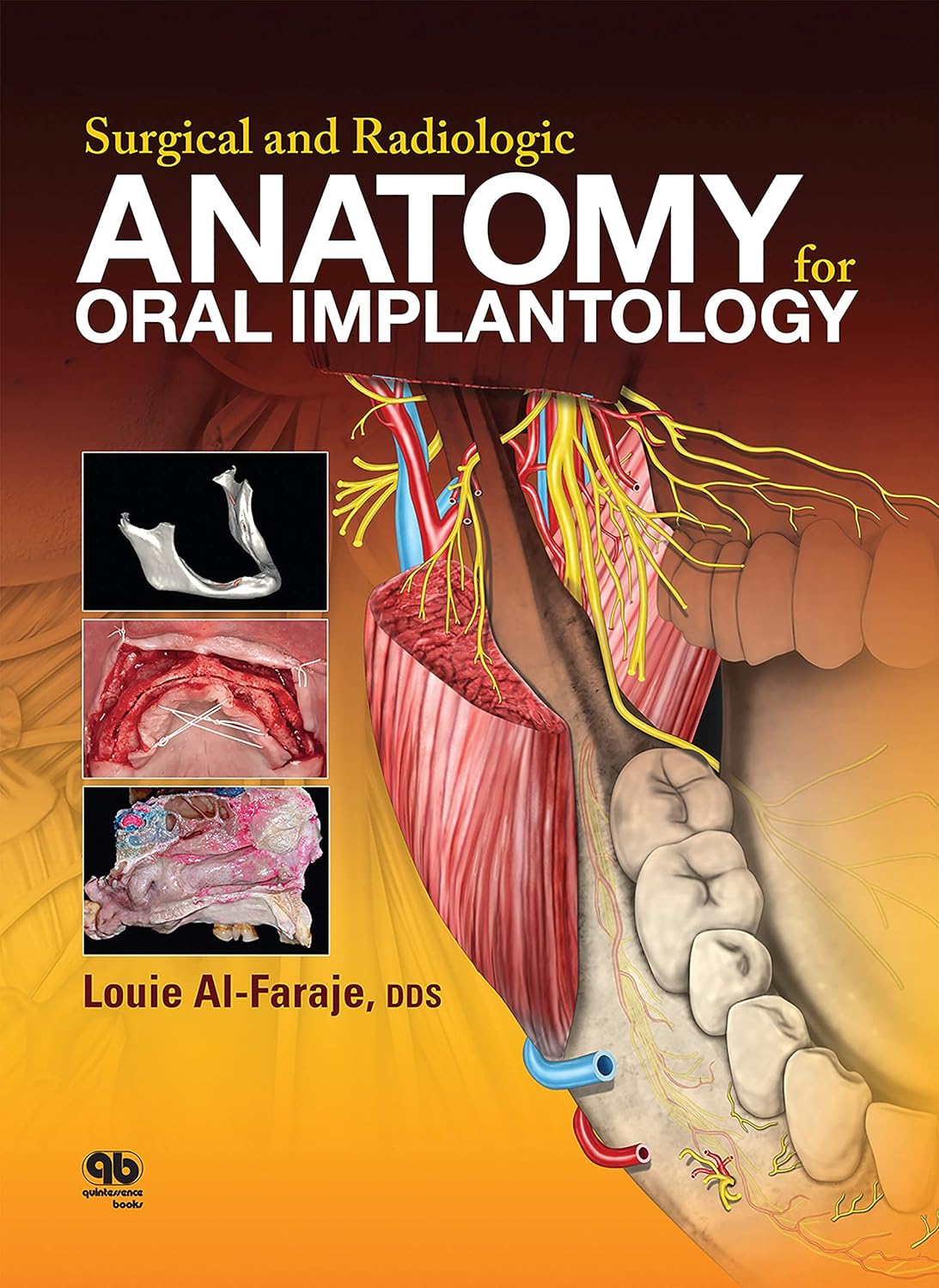
Clinical Anatomy for Oral Implantology – A Practical, Specimen-Based Guide
Clinical Anatomy for Oral Implantology provides a uniquely practical approach to the complex anatomy of the maxilla, mandible, and nasal cavity, tailored specifically for oral implantologists and dental surgeons. Unlike traditional anatomical texts, this book focuses on clinically relevant structures as they appear in cadavers and live patients, bridging the gap between theoretical knowledge and operative application.
Key Features
- Specimen-Based Learning: Uses cadaver dissections and clinical cases to depict anatomical structures as they exist in reality, avoiding confusion caused by overly schematic diagrams.
- Full-Page Labeled Images: Several chapters include detailed, full-page images of cadaver sections with clearly labeled structures for quick reference during study or surgery.
- Cone Beam CT Integration: Demonstrates the use of CBCT to measure bone density, alveolar ridge width, and distances to key anatomical landmarks, supporting precise implant planning.
- Surgical Relevance: Focused on simplifying the learning and execution of implant-related procedures in anatomically challenging regions.
- Bridging Theory and Practice: Helps both students and practicing clinicians gain a solid understanding of intraoperative anatomy for safer, more accurate implant placement.
This book is an essential resource for dental students, implantology trainees, and practicing clinicians seeking a practical, hands-on guide to the complex anatomy involved in oral implant surgery.