Skip to product information
1 of 1
🏷️ Save up to 80% on all courses, discount auto applied

Advanced functional dentistry in the concept of Mariano Rocabado

Advanced functional dentistry in the concept of Mariano Rocabado

Regular price $57.00 USD
Regular price $463.00 USD Sale price $57.00 USD
Sale Sold out
Shipping calculated at checkout.
Girl in a jacket

Instant Download

Your unique course download link appears right after you complete your purchase, and is also sent instantly to the email address you provided.

Professors

Roberto Gutierrez

Target audience

General dentists

Nb of lessons

13

Nb of modules

Course duration

16 h 51 min

Delivery method

Google Drive Link, Offline

Course Overview

Advanced training course on protocols for the treatment of craniovertebral, craniomandibular and TMJ dysfunction. Detailed analysis of protocols and clinical cases, covering the full aspect of rehabilitation of the listed above pathologies from A to Z awaits you.

During the course, you will learn:

  • Treatment planning according to the protocols of Dr. Mariano Rocabado
  • Protocols of radiological assessment of the TMJ and craniovertebral region
  • Splint therapy technique and total functional prosthetic rehabilitation
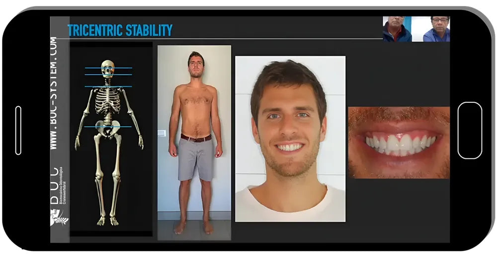
  • Tricentric Concept principles and their application in functional rehabilitation.

Training course from the legendary lecturer and the primary source of information, Mariano Rocabado!

You can't miss this!

Full Course Program

Lesson 1. Synovial temporomandibular joint integrative functional anatomy

  • The true definition of synovial joint
  • Anatomy of synovial temporomandibular joint
  • Anatomy of the temporal bone
  • Histological and anatomical analysis of the synovialmembrane
  • Craniocervical mandibular (CCM) tricentric relation
  • Determining the craniocervical position in relation to the CR
  • Radiological analysis of the TMJ.

Duration: 46 min · Presented by: Mariano Rocabado

Lesson 2. Differential diagnosis of tmd. analysis of clinical cases

  • Diagnosis of orofacial pain
  • Intraoral and extraoral diagnosis of TMD
  • Radiological assessment of the the TMJ
  • Cephalometric analysis and its significance in differentialdiagnosis
  • Differential diagnosis of TMD and orofacial pain
  • Treatment planning and analysis. Interdisciplinary approaches
  • Analysis of clinical cases.

Duration: 1 h 32 min · Presented by: Mariano Rocabado & Roberto Gutierrez

Lesson 3. Interdisciplinary approach. diagnosis and treatmentof tmd

  • Analyzing different treatment options
  • Limited mouth opening: diagnosis and treatment
  • TMJ hypermobility: diagnosis and treatment principles
  • Splint therapy
  • Craniovertebral and craniomandibular issues
  • Craniovertebral and TMJ dysfunction. Utilizing splint therapy
  • Combining splint therapy and orofacial myofunctional therapy(OMT)
  • Pivot splint: manufacturing and application features
  • Team approach: dentist-manual physician
  • Interdisciplinary approach to TMD. Splint therapy, orthodonticsand total functional rehabilitation
  • Functional wax-up of total functional rehabilitation cases
  • Step-by-step analysis of various clinical cases.

Duration: 1 h 23 min · Presented by: Roberto Gutierrez

Lesson 4. Vertical dimension of occlusion (vdo) in clinical practice

  • Anatomical, physiological and biomechanical considerationsof the craniovertebral region and the temporomandibular joint
  • Vertical dimension of occlusion in clinical practice
  • VDO in treatment of TMD
  • Tricentric stabilization
  • The effect of an increased and decreased VDO
  • Treatment of tooth wear
  • Analysis of various clinical cases.

Duration: 1 h 29 min · Presented by: Roberto Gutierrez

Lesson 5. Clinical anatomy of the synovial temporomandibular joint

  • Physiological anatomy of TMJ movement
  • CBCT analysis of the TMJ. Open and closed mouth
  • Evaluation of normal TMJ and clinical cases of TMD
  • Osteochondritis dissecans and avascular necrosis
  • CBCT and MRI considerations in various clinical cases.

Duration: 1 h 26 min · Presented by: Mariano Rocabado

Lesson 6. Cone beam computed tomography protocols of dr. mariano rocabado. Part 1

  • Analysis of cranioverteral according to Dr. Mariano Rocabado
  • Normal occiput atlas axis flexion
  • Craniofacial assymetry evalaution
  • CBCT and x-ray static and dynamic cervical and TMJ protocol
  • Functional analysis by Dr. Mariano Rocabado
  • Step-by-step of clinical analysis of various cases.

Duration: 54 min · Presented by: Mariano Rocabado

Lesson 7. Cone beam computed tomography protocols of dr. mariano rocabado. Part 2

  • Anatomy: what you need to know for the treatment of TM
  • Jpathology
  • Tricentric approach: advantages and special features
  • Map of joint pain: areas of sensitivity in the joint
  • Assessment of the state of the TMJ by CBCT
  • Static assessment of the spine from the side
  • Functional analysis by Dr. Mariano Rocabado
  • Craniocervical mandibular (CCM) tricentric relation
  • Step-by-step clinical analysis of various cases.

Duration: 54 min · Presented by: Mariano Rocabado

Lesson 8. Craniovertebral odontological biomechanics. Fundamentals of functional occlusion

  • Biological principles of occlusion
  • Occlusion pillars
  • Anterior and posterior vertical dimension
  • Complex tooth morphology
  • Anterior guidance. Canine guidance
  • Analysis of the curve of Spee
  • Evaluation of malocclusion. Functional considerations
  • The craniocervical mandibular (CCM) system
  • The pillars of stable occlusion
  • Centric relation and centric occlusion

Tricentric interactions:

  • Craniovertebral,Craniomandibular,Central occlusion at rest
  • Working with the articulator and mounting casts in CR
  • Application of the pivot splint
  • Functional waxing
  • Onlay restoration: from modeling to fixation
  • Analysis of clinical cases.

Duration: 2 h 14 min · Presented by: Roberto Gutierrez

Lesson 9. Craniovertebral radiology. a different point of view. Part 1

  • Body compensation and posture
  • Tricentric Concept
  • Parallel therapy: the dentist and physical therapist
  • Basic radiology concepts of cranio-vertebral region
  • TMD: disc and condyle position
  • The most common STMJ pathologies
  • Posture and head-mandible position.

Duration: 29 min · Presented by: María Fernanda Gutiérrez

Lesson 10. Craniovertebral radiology. a different point of view. Part 2TMJ anatomy. Pathological anomalies:

Degenerative process of the TMJ,Complete osteophyte,Erosion, flattening of both STMJ,Condylar post-surgical resorption,Condylar rheumatoid resorption,,Condylar hypoplasia,Condylar fracture;

Cervical anatomy. Development and pathological anomalies:

  • Signs and symptoms of cervical spine dysfunction
  • Cervical degenerative disc disease
  • Osteoarthritis of cervical vertebrae
  • Cervical herniated disc
  • Degenerative vertebral process
  • What is spondylolisthesis?
  • Craniovertebral and TMJ imaging protocols
  • Head position and its effect of the muscles of the neck
  • Analysis of clinical cases.

Duration: 1 h 31 min · Presented by: María Fernanda Gutiérrez

Lesson 11. The foundation of multidisciplinary treatment: dentist, physical therapist, speech therapist. Diagnosis and treatment planning

  • Analysis of occlusion
  • Biomechanics of the lower jaw
  • Posture and its effect on occlusion and treatment planning
  • Premature contact. Is it occlusal or craniovertebral?
  • Key to multidisciplinary treatment approach. Communicationand joint diagnosis
  • Functional rehabilitation cases
  • Working with the articulator: treatment planning protocols
  • Deprogramming splints: indications and clinical application
  • Analysis of clinical cases.

Duration: 1 h 06 min · Presented by: María Fernanda Gutiérrez

Lesson 12. The foundation of multidisciplinary treatment. Treatment protocols

Clinical and laboratory analysis of clinical cases:

  • Intra and extraoral diagnosis,Radiological assessment,Pivot splint therapy,Craniovertebral Orthopaedic Manual Therapy (COMT),Orthodontic treatment protocols,Functional rehabilitation protocols
  • Multidisciplinary approach in a clinical case: open bite withbilateral anterior and lateral disc luxation without reduction.

Duration: 1 h 55 min · Presented by: Mariano Rocabado & Roberto Gutierrez

Lesson 13. Photography and video protocol. Application in clinical practice

Protocols of the first appointment;

Protocols for documentation of clinical cases:
  • Intra and extraoral photography
  • Mandibular movement videos
  • Cervical movement videos
  • Pain zone photography
  • Analysis of clinical cases.

Duration: 1 h 12 min · Presented by: Mariano Rocabado & Roberto Gutierrez

View full details